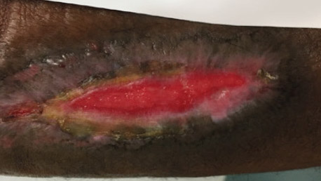
Case study: Necrotic eschar on right forearm
Wound type:
Necrotic eschar
Patient
69-year-old African-American male
History
Medical history of deep vein thrombosis, hip surgery, prostate cancer, ankle fusion, radial nerve palsy, and blood clots. Medications include warfarin, calcium supplements, and metoprolol. Non-smoker.
100%
granulated tissue achieved in 10 weeks*
*Individual results will vary
Wound presentation
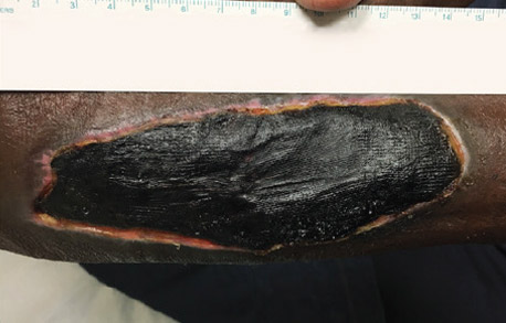
The hematoma was evacuated by an orthopedist in the emergency room, leaving full-thickness skin necrosis.
Treatment
Day 1
- Daily saline wash and application of SANTYL◊ Ointment, which facilitated a bloodless eschar debridement. Patient seen by physician every two weeks.
- Wound measures 13.1cm x 5.0cm
- Daily SANTYL Ointment application started by orthopedist three days prior to baseline and continued post-baseline
- After a daily wash, SANTYL Ointment was applied at home by patient on top and along
Day 28
Pre-debridement
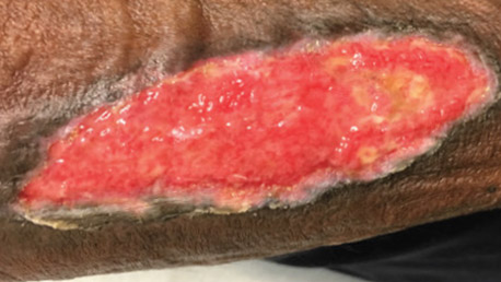
- Wound measures 10.4cm x 2.9cm, approx. 39% smaller from baseline
- No purulence, cellulitis, or odor
- Mostly granulation tissue with necrotic subcutaneous tissue visible throughout
- Wound is sharp debrided with a large curette removing the necrotic subcutaneous tissue
- Daily wash and SANTYL Ointment application with saline dressing continued
Day 56
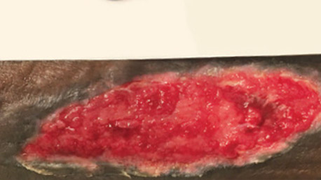
- Wound measures 5.2cm x 1.0cm, down 83% from Day 28 and 93% from baseline
- Surface area nearing complete granulation
- Some necrotic subcutaneous tissue
- No cellulitis
- Daily wash and SANTYL Ointment application with saline dressing continued
Result
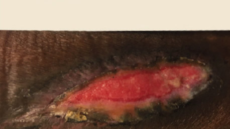
100%
granulated tissue achieved in 10 weeks*
*Individual results will vary
Download patient case study: Necrotic eschar on right forearm


