Case study: Diabetic ulcer on foot
Wound type:
Diabetic foot ulcer
Patient
89-year-old Caucasian male
History
Non–insulin-dependent diabetes mellitus (NIDDM)
Complete debridement achieved within four weeks*
*Individual results will vary
Wound presentation
- Ulcer on medial right hallux interphalangeal joint, noticed four days prior to first visit
- Seen by a primary care physician who prescribed an oral antibiotic
- Patient was applying Neosporin™ and a small adhesive bandage daily
Treatment
- Sharp debridement was performed at each visit
- Patient was instructed to wash and dry the wound daily
- SANTYL◊ Ointment was prescribed to be applied daily for debridement of necrotic tissue
- Dry gauze was applied because the ulcer had enough drainage to supply appropriate moisture balance
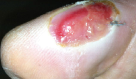
Day 1
- 2.3cm x 1.5cm; 0.2cm depth
- 20% red granular and 80% yellow fibrotic
- Mild sanguineous drainage
- No odor, no infection
- Sharp debridement performed
- SANTYL Ointment initiated
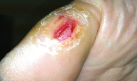
Day 16
- 0.8cm x 0.2cm; 0.2cm depth
- 85% red granular and 15% yellow fibrotic and hyperkeratotic margins
- Sharp debridement performed
- SANTYL Ointment continued
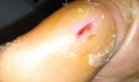
Day 30
- 0.5cm x 0.2cm; 0.1cm depth
- 100% red granular base and hyperkeratotic margins
- Sharp debridement performed
- SANTYL Ointment discontinued
Result
Complete debridement achieved within four weeks*
*Individual results will vary
Download patient case study: Diabetic ulcer on foot


