Case study: Chronic leg wounds
Wound type:
General Wound
Patient
60-year-old female
History
Chronic leg wounds for two years. Non-compliant with a prior wound care center and transferred for plastic surgery flap coverage. The patient’s past medical history includes sickle cell disease with a baseline Hb6 and mild venous stasis disease. The patie
100%
re-epithelization on day 42*
*Individual results will vary
Wound presentation
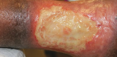
Two distinct, heavily exudative wound ulcers; one on the right medial ankle and one on the right lateral distal ankle. There was no initial trauma of the leg reported and the ulcers were consistent with chronic sickle cell disease.
Treatment
- No treatment for three months prior to the wound presentation outside of dressing changes
- Once initiated, treatment included sharp debridement followed by SANTYL◊ Ointment application with xeroform covering and compressive dressing changes once daily for 35 days
Day 1
- Right lateral ulcer measures 10.0cm x 8.0cm; 0.5cm depth
- Right medial ulcer measures 10.0cm x 10.0cm; 0.5cm depth
- No granulation tissue or gross infection; 100% slough noted
- MRI results negative for deep space collection or osteomyelitis
- Sharp debridement performed
- Daily SANTYL application with xeroform covering and compressive dressing changes initiated
Day 14
- No change in wound dimensions from baseline
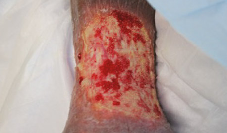
- Improvement in overall wound appearances; 20% granulation tissue throughout both wounds
- Ongoing serous drainage and slough noted on both wounds
- Decreased swelling in leg
- Sharp debridement performed
- Daily SANTYL Ointment application with xeroform covering and compressive dressing changes continued
Day 28
- No change in wound dimensions from baseline
- Continued improvement noted; 80% granulation tissue present throughout both wounds
- No evidence of infection
- Sharp debridement performed
- Daily SANTYL Ointment application with xeroform covering and compressive dressing changes continued
Day 35
- No photos available for day 35
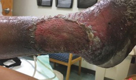
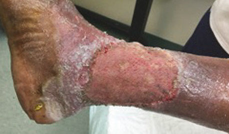
- Post-skin graft
- Wound VAC dressing cover removed
- Graft healed to 100% re-epithelization
- Staples removed
- Moisturizer and ongoing compression prescribed
- Patient discharged to primary follow-up team
Day 56
- Continued improvement noted; 100% granulation
- Minimal slough
- SANTYL Ointment discontinued
- Sharp debridement and split-thickness graft application with wound
- VAC dressing cover performed in operating room
Result
100%
re-epithelization on day 42*
*Individual results will vary
Download patient case study: Chronic leg wounds


